A Complex Extraction and Implant Case Involving the Maxillary Sinus
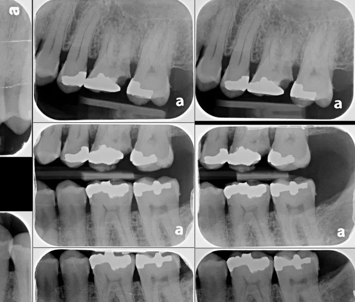
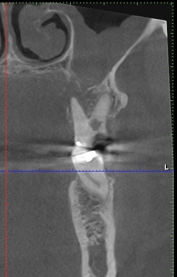
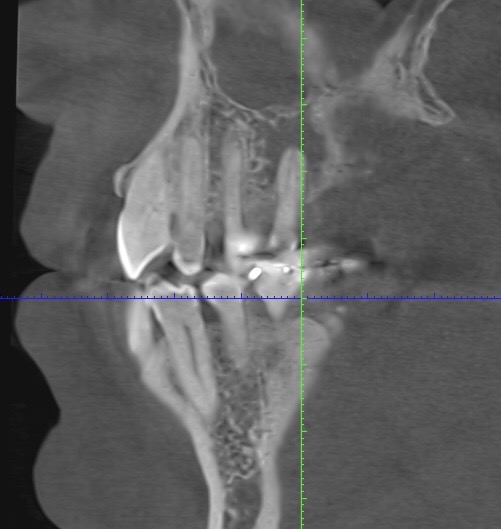
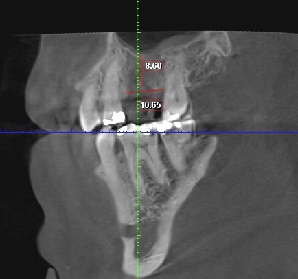
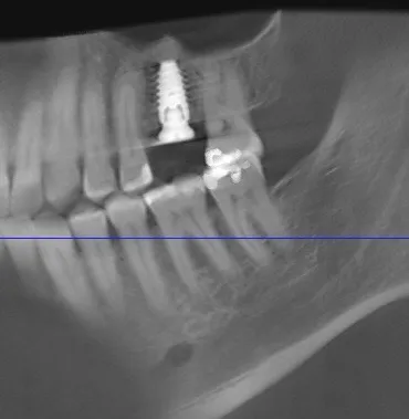
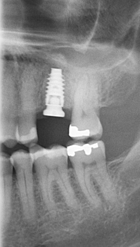
It amazes us how well people can heal sometimes. This case, done by Dr. Wes Parker, involved a 45 year old male who was referred for a carious, necrotic (dead) tooth #14 (upper left first molar). The patient also reported left sided sinus pressure and drainage. At his consultation, we obtained a Cone Beam CT (CBCT) scan. On his CBCT, you could see where the infection and inflammation from tooth #14 had eroded through the floor of the left maxillary sinus. We discussed this with him and scheduled surgery shortly after the consultation. Dr. Parker removed tooth #14, cleaned out the pus and inflamed maxillary sinus lining through the socket. The inflamed, thickened sinus lining went almost up to the orbit (eye socket). Dr. Parker then closed off the sinus communication. Following this, Dr. Parker placed a bone graft over the sinus closure, and then sutured over that to close the wound. The patient healed very well. He was compliant with the sinus precautions and medications that were prescribed. Next, Dr. Parker proceeded with implant placement in the #14 site with a simultaneous indirect sinus lift or “sinus bump.” The implant placement surgery went well, and after about 5 months of healing time, the patient received a crown on the implant from his dentist. Image #1 is a preoperative PA radiograph of the carious, necrotic, nonrestorable tooth #14 (upper left first molar). Image #2 is a coronal slice from the patient’s preoperative CBCT showing the necrotic tooth #14 with a periapical radiolucency and maxillary sinus inflammation stemming from the necrotic tooth. Image #3 is a sagittal slice from the patient’s preoperative CBCT showing the necrotic tooth #14 with a periapical radiolucency and maxillary sinus inflammation stemming from the necrotic tooth. Image #4 is a sagittal CBCT slice from the patient’s post extraction CBCT showing resolution of the sinus inflammation and measurements for the future dental implant. Image #5 is an immediate postoperative PA of the implant in the #14 site following implant placement with a simultaneous indirect sinus lift. Image #6 is a 5 month postoperative PA showing the healed, integrated implant prior to crown placement with a healed sinus lift bone graft above it and no sinus inflammation.